


State of the Art Lab
Central to any consideration of techniques in the IVF lab is the quality of the gametes that the lab has to work with. Usually there is little opportunity to modify the basic quality of sperm, however, the oocyte is a direct biological end product of ovarian super-ovulation as commonly conducted in human IVF therapy.
Objectives:
- present current choices for embryo transfer at different stages.
- discuss strategies for embryo cryopreservation at different developmental stages.
- consider technologies thought to enhance embryonic implantation.
- look at new technologies that may gain a foothold in the ART lab.
Central to any consideration of techniques in the IVF lab is the quality of the gametes that the lab has to work with. Usually there is little opportunity to modify the basic quality of sperm, however, the oocyte is a direct biological end product of ovarian super-ovulation as commonly conducted in human IVF therapy. The introduction of recombinant technology to the production of oocytes has finally brought us to a point where, assuming stimulation is conducted appropriately, that oocyte quality can now be considered to have a direct relationship with the fertility status of the woman. Once collected, both oocytes and sperm enter the artificial environment of the in vitro laboratory. What is done thereafter to produce and influence embryo quality and potential for pregnancy is a constant theme of the ART laboratory.
In vitro culture.
Moving from an era when IVF culture media were mostly simple basic salt solutions and other concoctions not specifically designed for human gametes and embryos, Quinn et al. (1985) were the first to attempt to mimic physiological conditions in the Fallopian tube by matching principally the potassium/sodium ion concentrations, this being the basis of the medium known as HTF ("human tubal fluid"). Since that time the logical conclusion of this approach has been explored in the form of co-culture, where a complete quasi-in vivo environment is created to replicate either the uterine or the tubal milieu (Wiemer et al 1998). The demise of co-culture has been the drive to more fully define the in vitro conditions to increase consistency of in vitro culture. This set the stage for embryo stage specific media to be designed (Gardner & Lane, 1998), matching conditions in vitro with those in vivo all the way to the blastocyst stage. Such an equivalence theory has merit, but relies on certain assumptions that the physical environment is identical between the body and the culture dish. It may be that conditions in vitro are so "artificial" that in vitro culture requirements differ for the embryo in these circumstances. Ultimately, the success of a culture medium is probably still principally driven by the quality of its ingredients, not just by the specific formulation. Recently improved QC conditions of media manufacture have gone far to improve overall IVF outcomes by consideration of this fact. Indeed, autologous cell co-culture (Freeman et al, 1995) might still have relevance for example, in overcoming the artifice of in vitro culture. Alternative systems where the physical conditions of in vitro culture are dramatically changed, may provide a more appropriate environment for in vitro embryos, e.g., hyperbaric, "submarine" culture with constant agitation of the embryos in their sealed dishes (Anita Sjgren & Lars Hamberger, Personal Communication).
How to culture gametes and embryos remains an issue of debate, though use of oil overlay to optimize culture volumes and conditions is increasingly recognized, with some groups placing faith in actual co-incubation of sibling embryos (Lane et al, 1992). More recently the merits of group culture of human embryos, at least through early cleavage, have not been confirmed (Spyropoulou et al. 1999). The need to culture successfully human embryos for longer periods of their development is becoming increasingly clear, even though normal pronucleate uterine transfer can yield results (Scott & Smith, 1998). Increased culture periods, however, increase the level of embryo selection, to maximize the choice of the best quality embryos for fresh transfer, and to minimize the need for transfer of greater numbers of embryos; i.e., to optimize IVF success through increased pregnancy rates, while reducing multiple implantation (Elsner et al, 1997).
The simple expedient of extending culture to day-3 of development versus day-2 has improved results over the past 8 years. The time has now come (Jones et al, 1998)) to push that period out routinely to day-5/6 to allow embryos to "select" themselves by growth to blastocysts. It would be wrong to suggest that all blastocysts are equal in viability, and experience is rapidly being gained in better defining blastocyst quality in terms of morphology and development. Practical considerations exist though, such as the need possibly to double incubator space due to having embryos in the lab longer. Counselling of couples will have to incorporate an appreciation that chances for transfer and the number of embryos for transfer and cryostorage will be reduced. Indeed, clinics may have guidelines of how many cleaving embryos a couple might have to justify extended culture for increased embryo selection - a possible cut off might be five. Some poor prognosis couples with repeated failure of IVF-ET with minimal embryo numbers might seek blastocyst culture as a diagnostic tool, to see if their embryo have the wherewithal to grow to this stage prior to any further transfers. If this were not the case then such a couple might use this outcome to bring to a close IVF therapy for their infertility and move on with their lives.
Most clinics have either begun to address, or will soon consider the introduction of blastocyst culture as a greater or lesser adjunct to their IVF programs. The issue is how to do this most effectively. A clinical niche in which blastocyst transfer may readily be introduced could be an oocyte donation program, where potential for more and higher quality oocytes would improve potential for routine blastocyst outgrowth. By extension of this idea, the first couples that might be exposed to this approach with their own gametes could be those with the best prognosis, and at highest risk for multiple implantations following multiple embryo transfer. While these areas of therapy will yield the very best outcomes in terms of pregnancies following blastocyst transfer, the true aim is to minimize transfers of more than two embryos to limit multiple pregnancies mostly to twins only. Moving to poorer prognosis couples that say have had multiple failed IVF-ET cycles, each time with a fair selection of embryos for transfer or cryopreservation on day-three of development. Such couples could well benefit from extended culture to truly establish which embryos are of the highest viability. This may mean that embryos do not get utilized at such a high rate as on day-three, but will generate a clearer picture of how "selected" embryos on day-three ultimately grow out to blastocyst. Some of these couples may have no blastocysts form and may not even have an ET. This may seem brutal, but assuming one has confidence in the in vitro culture system itself, then such outcomes can be seen to be diagnostic.
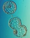
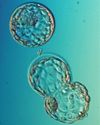
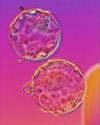
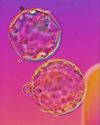
In vitro grown human blastocysts on day-five of development,
either hatching or about to hatch free from their zonae pellucidae

Experience with Clinical Blastocyst Use at Shady Grove Fertility Centers, Maryland.
As an illustration of some of the above points, it might be helpful to describe the experience of the introduction of routine blastocyst culture at Shady Grove Fertility Centers under the diligent guidance of Jim Graham at that facility. Using 15-35 microliter droplets of medium under mineral oil as the standard culture approach, embryos were individually cultured, and a three stage culture medium system of HTF and Basal XI media (HSA suppplemented) for day-0 through day-3 was utilized, with a direct change over into S-2 medium late on day-3 for blastocyst culture. [More recently the full range of HTF/G1.2/G2.2 has been utilized with comparable results]. Blastocysts were anticipated either day-5 or day-6 with an estimate of when this might be, based on growth through the first three days. This change in routine IVF-ET protocol required a greater degree of flexibility on the part of the nursing and clinical staff, which necessitated changes in communication, patient counseling and expectations. Simple questions of when to begin progesterone supplementation, time of ET and pregnancy test all required some significant adjustment outside of the IVF lab itself.
From the lab perspective, it was of immense interest to correlate traditional estimates of embryo quality judged by rate of development and morphology on day-three, with what actually occurred on day-5/6. Surprisingly there was significant disparity between the two stages in embryo viability estimates. Analysis of traditional parameters for selection of embryos for fresh ET or cryopreservation on day-3 were superseded by extended culture and selection of blastocysts 2 or 3 days later. Efficiency of embryo selection for potential viability at both stages was compared by correlating individual embryo quality assessment as a proportion of assumed good quality embryos in both circumstances and ultimate clinical outcome. In 101 IVF cycles where women (average age 33.1 years) had sufficient embryos (range 5-25) to justify some degree of embryo selection, extended in vitro culture was undertaken for an average of 5.4 days before use. From 1263 cleaving embryos 559 were judged to be of a standard suitable for use on day-3 by traditional parameters of selection. Day-3 use would have dictated that 355 embryos would have been transferred (average 3.5) and the remainder frozen. This would have been a 44% utilization of all cleaving embryos (559/1263) at that time. This compares with 234 blastocysts that were actually transferred (average 2.3) and 237 that were cryopreserved, where embryo utilization dropped to 37% of all cleaving embryos. This rate differs from "blastocyst formation rate", commonly quoted as about 45-50%, which does not account for quality of the embryo and whether it was actually used for ET or cryopreservation. Interestingly, by following individual embryo culture it was possible to compare the original embryo's potential use for ET, cryopreservation or discard through to its actual fate as a blastocyst. These data show that at Shady Grove only 47.5% (265/559) of day-3 embryos that would have been considered suitable for ET or cryopreservation were ultimately chosen for ET or cryopreservation as blastocysts. Additionally, 206 day-3 embryos that would not have been chosen for use on day-3, were used as blastocysts. Of day-3 embryos that would have been transferred, tellingly only 39% would be transferred fresh as blastocysts; not a very resounding testament to traditional selection criteria on day-3!
It may not be ideal, but an historic comparison of ongoing pregnancy rates per transfer in the Shady Grove program before and after the adoption of blastocyst culture for fresh transfer indicates an effective jump from 30.5% for day-3 transfers to 45% for day-5/6 transfers in comparable patients. A conservative "guesstimate" of overall program improvement with blastocysts transfers as routine would, in addition to reducing high order multiple pregnancies, perhaps see a rise in pregnancy rates of between 5-10% per transfer. The number of cases that would fail to achieve transfer would reflect the clinic's patient selection criteria, but could be as low as only 5-7% of all egg collections initiated.
Table 1: Patient and IVF cycle breakdown
101 (average age 33.1 years; range 24 - 44)
Table 2: Day-3 embryo and blastocyst "selection" comparison
Cryostorage of the female gamete.
The last few years have seen a significant resurgence of interest in the potential benefits of human egg freezing. Essentially, these benefits are:
- Formation of donor "egg banks" to facilitate and lessen the cost of oocyte donation for women that are unable to produce their own oocytes.
- Provision of egg cryostorage for women wishing to delay their reproductive choices.
- Convenient cryopreservation of ovarian tissue taken from women about to undergo cancer therapy, which may threaten their reproductive health.
The technology so far applied clinically has been based directly on traditional human embryo cryopreservation protocols, and has produced relatively few offspring. Fortunately to date, no abnormalities have been reported from these pregnancies, regardless of the persistent concerns that freezing and thawing of mature oocytes may disrupt the meiotic spindle and thus increase the potential for aneuploidy in the embryos arising from such eggs. With respect to cryo-storage of donated oocytes there have been three centers Worldwide that have so far reported some degree of success with this approach (Polak de Fried et al, 1998; Tucker et al, 1998a; Yang et al, 1998 & personal communication). Six ongoing/delivered pregnancies have generated approximately 10 viable fetuses or delivered babies from cryopreserved donor oocytes. Use of frozen donor oocytes post-thaw not for whole egg donation but for ooplasmic transfer has been reported with a successful delivery of a twin following thawed ooplasmic donation (Lanzendorf et al., 1999).
Cryostorage of women's own oocytes was originally reported in the case of three births over a decade ago by two centers (Chen, 1988; Van Uem et al, 1987). More recently these successes have been reproduced by others (Porcu et al, 1998; Tucker et al, 1998b; Yang et al, 1998 & personal communication), giving rise to eight ongoing/delivered pregnancies with 9 viable fetuses or delivered babies. One other baby has arisen from a clinical circumstance that is not completely unfamiliar to IVF clinics, where oocytes have been collected but the partner was unable to produce spermatozoa available for insemination. In just such an instance, oocytes were frozen, and spermatozoa were collected and frozen at a later date. Then ultimately both sets of gametes were thawed and used in a subsequent IVF attempt, which achieved a health delivery (Moody & San Roman, personal communication).
All of these pregnancies were from frozen-thawed mature oocytes, but for one notable exception, where a pregnancy arose from an immature germinal vesicle (GV) stage egg (Tucker et al, 1998b). Interestingly, this stage of egg development may prove to be a more successful approach for cryopreservation because its oolemma is more permeable to cryoprotectant, and its chromatin is more conveniently and safely packaged in the nucleus (Van Blerkom & Davis, 1994). Such eggs, however, still have to undergo GV breakdown and maturation to the MII stage before fertilization, and therefore their developmental competency is not so clearly established as with fully mature oocytes that are frozen. Source of the GV eggs and whether they have been exposed to any exogenous gonadotropins may play a key role in the competency of these eggs (Cortvrindt et al, 1998).
Whether mature or not, standard cryopreservation technologies appear to have their ultimate limitations in not only cryosurvival, but also more importantly in their lack of consistency. 50% cryosurvival is an adequate overall outcome, but not if it is a statistic that is arrived at by 90-100% survival in one case, and 0-10% in the next. Consequently, radically different types of protocol may provide the answer to increased consistent success. One approach has been to replace sodium as the principal cation in the cryoprotectant with choline in an attempt to shut down the sodium ion pumps in the oocyte membrane during cryoprotectant exposure, thus minimising potentially deleterious "solution effects" during cooling (Stachecki et al, 1998). This has provided significant improvements in murine egg freezing, and though not yet applied clinically in the human, it may be a promising advance. Alternatively, traditional slow cooling/rapid thaw protocols might be replaced with vitrification. Which again has been successfully applied in the mouse (O'Neil et al, 1997).
The most plentiful source of oocytes potentially is ovarian tissue itself, containing as it does many thousands of primordial follicles in healthy cortical tissue. Earlier successful work with cryopreservation of rodent ovarian tissue has led the way to successful cryostorage of both sheep and human tissue (Gosden et al, 1998). Up to 80% survival of follicles has been reported, but the issue is how to handle this tissue following its thaw. Tissue that has been removed, for example, from a woman about to undergo cancer therapy may contain malignant cells, and therefore may not be safely used for autografting in to such a woman if she were to survive. The tissue might be screened before or after thawing for the presence of malignant cells to enable some assessment of the safety of such an approach, or it may be grown in a host animal (e.g., SCID mouse) until such time as in vitro maturation could be undertaken more effectively. Extended culture of primordial follicles to full oocyte maturity, with subsequent embryonic development and birth has only been recorded in the mouse, and this was not from cryopreserved tissue (Eppig & O'Brien, 1996). Fertility has been restored in sheep, in a good model for the human ovary, following cryostorage of ovarian cortex and autografting (Gosden et al, 1994), and this seems the most likely successful clinical model for restoration of fertility of women who are at risk of losing their ovarian function. This may include not only women about to undergo cancer therapy, but also women who have a family history of early menopause, and those with non-malignant diseases such as thalassemia or certain auto-immune conditions which may be treated by high-dose chemotherapy.
The very many potential routes for cryostorage of the female gamete makes for a confusing vision of where clinical applications may occur. However, different clinical needs may actually be met by differing technological approaches, whether they incorporate whole tissue freezing, separate follicle storage, or cryopreservation of mature oocytes themselves.
Cryopreservation of the preimplantation human embryo.
While human embryo cryopreservation has become a well-established technology in assisted human reproduction, it has yet to become fully clear as to which stage preimplantation embryos are best cryostored. Indeed on the face of it, the superiority of blastocyst stage freezing over 1-cell pronucleate stage freezing in terms of implantation per thawed embryo transferred, is countered by the loss of embryos that lack the wherewithal to grow for 5 to 6 days in vitro (Mandelbaum et al, 1998). Thus there seems to remain some degree of clinic choice of philosophy of approach over when to freeze (Tucker et al, 1995). If one is to assume, however, that the majority of in vitro culture of human embryos will eventually be carried out to the blastocyst stage, then it would seem redundant to freeze embryos at an earlier stage. Not to belabor the point, but selection is the central essence of extended culture, enabling poorer viability embryos to arrest in development so "selecting" themselves as non-candidates for fresh transfer or cryopreservation. Although to some this may seem wasteful of embryos, the net result is probably that chances of pregnancy are more clearly defined and potentially improved, whilst the risks of higher order multiple implantations are reduced. Additionally, fewer embryos will be frozen as blastocysts, reducing storage requirements, and one hopes, expectations of pregnancy of those embryos that are frozen will be improved. Therefore, overall efficiency will be increased.
Nevertheless, given the consistently high rates of cryosurvival of cryopreserved early stage embryos, there will probably continue to exist certain clinical circumstances where early stage freezing is justified. Indeed, there remain some clinics that continue to hone their abilities to achieve high rates of success with study of the culture conditions of pronucleate embryos post-thaw (Mayer, personal communication). Regardless of wishing to move a clinic's cryopreservation program to blastocyst stage only, a key question is what to do with those early stage embryos already cryostored. One progressive approach may be to thaw all embryos at these earlier stages and grow them to blastocysts if possible. In this way, fewer embryos will be kept cryostored, and if an excess of embryos for transfer do reach the blastocyst stage, then they may be re-frozen for later use.
Although the first successful reports of human blastocyst cryopreservation came from culture in a simple salt solution (Fehilly et al, 1985), but more recently most cryopreserved blastocysts arose from extended culture of supernumerary embryos not transferred fresh on day-2 or 3, usually using co-culture (Kaufman et al, 1995; Freeman, 1998). However, with increased confidence in growth stage-sequenced culture media, blastocyst culture for fresh transfer has become increasingly common. More convenient cryopreservation protocols for blastocysts (Menezo & Veiga, 1997) have also improved the ease with which this adjustment in a clinic's protocols may be made. Consistently high cryosurvival rates (approximately 90%), and good post-thaw pregnancy rates (38%) are now being achieved by certain clinics with judicious selection of blastocysts for freezing (Marek & Langley, personal communication). The key is how to select potentially viable blastocysts. As culture is extended over a longer period, rate of development becomes an increasingly important parameter for blastocyst selection (Shoukir et al, 1998); although a range of selection criteria need to be applied to optimize the choice of the blastocysts with the best potential for successful cryopreservation (see below).
Selection Criteria for Human Blastocysts for Cryopreservation:
- Expanded blastocyst growth rate: day-5 > day-6 > day-7
- Overall cell number > 60 cells (depending on day of development)
- Relative cell allocation to trophectoderm / inner cell mass
- Original quality of early stage embryo: PN formation, blastomere regularity, mononucleation, fragmentation
Issues such as how "early" a blastocyst can be frozen, or if blastocysts that are partially or totally hatched can be consistently cryopreserved, have yet to be adequately answered. Much data may exist from mouse and bovine models, for example, however cell number and levels of lipidation may have a profound differential impact thus minimizing the usefulness of such comparative studies. Hence, data will be collected as has often been the case with human ART prospectively, and used to fine tune future protocols from clinical hindsight. Most embryo cryopreservation protocols currently used, employ a slow freeze / rapid thaw approach. The more convenient protocols of ultra-rapid freezing and vitrification, that eliminate the use of expensive controlled rate freezers, await cross over from use in other species, or validation from more extensive experimental study in humans (Vatja et al, 1998; Lai et al, 1996). Regardless of such uncertainties, the future seems to point to increasing success and consistency with embryo cryopreservation. The preparation of the uterus into which the thawed embryos will ultimately be placed seems to be an area of study that is better resolved, with both natural and hormone replacement cycles providing comparable levels of receptivity in naturally cycling women, though differing levels of convenience (Tucker et al, 1995a). Indeed, artificially prepared cycles may even effectively dispense with the use of GnRH-a to lessen cost and improve convenience without loss of success (Simon et al, 1998).
In vitro maturation.
Reports of immature oocyte retrieval from unstimulated ovaries exist (Barnes et al, 1995; Cha et al, 1996). These immature occytes can successfully be cultured to the MII stage in vitro to achieve live births. The overall efficiency of this approach, however, remains in question. Some degree of ovarian stimulation will inevitably increase the potential for recruitment of more competent oocytes, and even if GV oocytes are sought for freezing or nuclear transfer, stimulation protocols tailored to maximize collection of such eggs are to be favored. In vitro culture of ovarian tissue itself following cryostorage (Oktay et al, 1998), may prove to be an effective means to reconstitute a woman's fertility after successful cancer therapy, for example, without the need to autograft ovarian tissue with its risk of reseeding the woman's body with cancerous cells (Gosden et al, 1997).
Micromanipulation.
Assisted Fertilization
ICSI (Palermo et al, 1992) has not only become the insemination of choice for all cases of IVF where fertilization failure may occur, indeed, it may well become the only form of insemination in vitro (Tucker et al, 1995b; Miller KF et al, 1998).
Assisted Hatching
This technology continues to be applied with varying reports of effectiveness (Schoolcraft et al, 1994; Lanzendorf et al, 1998). When undertaken with acidified Tyrode's medium to drill the zona pellucida, fragment removal can be carried out with some reported success (Alikani et al, 1999). Laser zona ablation (Antinori et al, 1996 ) may be also used, as well as physical rupture of the zona with a glass needle (Cohen et al, 1990). Assisted hatching could well be eliminated in its present forms to be replaced by whole zona dissolution prior to blastocyst transfer (Jones et al. 1998), as we learn more of the actual characteristics of zona pellucida behavior under the influence of the expanding blastocyst, and how this might correlate with the timing and potential of hatching. The risk of exposure of the "naked" blastocyst and its trophectoderm to the enzyme pronase and its vulnerability during ET, has to be weighed against the potential benefits of releasing the blastocyst from all zonal constraints during its preimplantation phase.
Again to use experience at Shady Grove Fertility Centers in the past six months, it was found that there was a distinct difference in ongoing pregnancy and implantation rates between day-5 and day-6 transfers: Preg.Rate 56% vs. 25%; Emb. Imp. 33% vs. 12%. The simplistic interpretation would be that day-6 transfers are "bad". However, in fairness the patients in this group (n=41) were older and had undergone more repeat cycles than the day-5 cases (n=67). In an attempt to address this poorer outcome, we undertook assisted hatching with acid Tyrode's on all day-6 transfer cases following exposure of the blastocysts to 0.1M sucrose to minimize the risk of damage to the trophectoderm during drilling. To date of 28 cases, 13 (46.5%) now have clinical pregnancies, an apparently distinct improvement in outcome in these cases. A large hole can be drilled (30-40microns) with little risk of disruption to the blastocyst, and commonly hatching is observed at the time of transfer. A laser would be very convenient for this process. It is probable that routine hatching of thawed blastocysts may be beneficial, drilling the zonae before re-expansion, thus avoiding the need for sucrose shrinkage of the blastocyst.
Embryo Biopsy
As the ability to obtain more and more feedback on the normality and viability of an embryo from a single cell grows, then clinical justification for this technology will grow apace. Currently polar body biopsy from the oocyte, and blastomere biopsy from cleavage stage embryos has been applied successfully to screen for embryo sex, aneuploidy, translocations, and single gene region disorders (Verlinsky, 1996; Munn et al, 1995; Munn et al, 1998; Harper, 1996). Trophectoderm biopsy from the blastocyst has yet to prove to be so popular an approach due mostly to the shorter period of time available between biopsy and the time of transfer, limiting the time available to run a diagnostic screen. Though this problem is countered in part by the greater number of cells available for removal. Coupling of laser zona ablation with biopsy (Montag et al, 1998; Veiga et al, 1997) will facilitate removal of tissue.
Cytoplasmic and Nuclear Transfer
Cytoplasmic transfer has been applied successfully (Cohen et al, 1998); nuclear transfer has not. Both technologies, in essence, attempt to address the same problem: that of reduced viability of embryos in certain poor prognosis and older patients. Oocyte donation, or indeed embryo donation can be utilized to overcome apparent or total inability to produce viable embryos for some couples. Cytoplasmic transfer, on the other hand, attempts to bolster egg and subsequent embryo quality through injection of donor egg cytoplasm to a recipient's own eggs.
Nuclear transfer takes this a step farther by replacing all of the recipient's ooplasm with donor egg cytoplasm, having enucleated a donated egg for the purpose of transferring the patient's own nucleus in to that cytoplasm (Zhang et al, 1999; Takeuchi et al, 1999). The hope is that nuclear transfer at the GV stage would allow more successful meiosis to occur with the patient's own chromatin, within a younger healthier egg cytoskeleton.
Concluding Remarks.
All of these technologies considered, regardless of their ultimate clinical relevance, would not have reached or come close to reaching clinical application were it not for the skills and perseverance of clinical embryologists working within the practical constraints of human infertility therapy. While it would be wrong not to acknowledge the need for a successful team approach to ART, nevertheless it would be foolhardy not to support the scientific staff through investment in equipment and well-scrutinized experimental protocols. Such investment will bring us all closer to our goals of safer and yet more successful infertility programs.
References:
References
1. Al-Hasani S et al (1998) Comparison of cryopreservation of supernumerary pronuclear human oocytes obtained after intracytoplasmic sperm injection and after conventional in-vitro fertilization. Hum Reprod 11:604-7.
2. Alikani M et al (1999) Human embryo fragmentation in vitro and its implications for pregnancy and implantation. Fertil Steril 71:836-42.
3. Antinori S et al (1996) Zona opening of human embryos using a non-contact UV laser for assisted hatching in patients with poor prognosis of pregnancy. Hum Reprod 11:2488-92.
4. Barnes F et al (1995) Blastocyst development and birth after in vitro maturation of human primary oocytes, intracytoplasmic sperm injection and assisted hatching: a case report. Hum Reprod 10:3243-7.
5. Cha KY et al (1996) Successful in vitro maturation, fertliization and pregnancy by using immature follicular oocytes collected from unstimulated polycystic ovarian syndrome patients. Proc 52nd Ann Mtg ASRM, Boston, Abstr 0-044.
6. Chen C, 1988: Pregnancies after human oocyte cryopreservation. Ann NY Acad Sci 541: 541-549.
7. Cohen et al (1990) Impairment of the hatching process following IVF in the human and improvement of implantation by assisted hatching using micromanipulation. Hum Reprod 5:7-13.
8. Cohen J et al (1998) Ooplasmic transfer in mature human oocytes. Molec Hum Reprod 4:269-80.
9. Cortvrindt RG et al (1998) Timed analysis of the nuclear maturation of oocytes in early preantral mouse follicle culture supplemented with recombinant gonadotropin. Fertil Steril 70:1114-25.
10. Dulioust E et al (1995) Long-term effects of embryo freezing in mice. PNAS, USA 92:589-93.
11. Elsner CW et al (1997) Multiple pregnancy rate and embryo number transferred during in vitro fertilization. Am J Obstet Gynecol 177:350-7.
12. Eppig JJ & O'Brien MJ (1996) Development in mouse oocytes from primordial follicles. Biol Reprod 54:197-201.
13. Fehilly CB et al (1985) Cryopreservation of cleaving embryos and expanded blastocysts in the human: a comparative study. Fertil Steril 44:638-644.
14. Freeman MR (1998) Culture and selection of blastocysts for cryopreservation. Course II Postgraduate Program ASRM, San Francisco, Oct3-4 1998, pp107-127.
15. Gardner DK & Lane M (1998) Culture of viable human blastocysts in defined sequential serum-free media. Hum Reprod 13 (supplement 1):101-12.
16. Gook DA et al (1995) Intracytoplasmic sperm injection and embryo development of human oocytes cryopreserved using 1,2-propanediol. Hum Reprod 10:2637-41.
17. Gosden RG et al (1994) Restoration of fertility to oophorectomised sheep by ovarian autografts stored at -196oC. Hum Reprod 9:597-603.
18. Gosden R et al (1998) The cryopreservation of human ovarian tissue. Proc XVI World Congr Fertil Steril, San Francisco, 4-9 Oct 1998, Eds: Kempers RD, Cohen J, Haney AF, Younger JB, pp615-620.
19. Harper JC (1996) Preimplantation diagnosis of inherited disease by embryo biopsy: an update of the world figures. JARG 13:90-5.
20. Jones GM et al (1998) Evolution of a culture protocol for successful blastocyst development and pregnancy. Hum Reprod 13:169-77.
21. Kaufman RA et al (1995) Cocultured blastocyst cryopreservation: experience of more than 500 transfer cycles. Fertil Steril 64:1125-9.
22. Lai AC et al (1996) Pregnancies after transfer of ultrarapidly frozen human embryos. J Assist Reprod Genet 13:625-628.
23. Lane M et al (1992) Effect of incubation volume and embryo density on the development and viability of preimplantation mouse embryo in vitro. Hum Reprod 7:558-62.
24. Lanzendorf SE et al (1998) A prospective, randomized, double-blind study for the evaluation of assisted hatching in patients with advanced maternal age. Hum Reprod 13:409-13.
25. Lanzendorf SE et al (1999) Pregnancy following transfer of ooplasm from cryopreserved -thawed donor oocytes into recipient oocytes. Fertil Steril 71: In Press.
26. Mandelbaum J et al (1998) Cryopreservation in human assisted reproduction is now routine for embryos but remains a research procedure for oocytes. Hum Reprod 13; suppl 3, June 1998, pp161-174.
27. Ménézo Y & Veiga A (1997) Cryopreservation of blastocysts. Proc X World Congr IVF and Assisted Reprod, Vancouver, 24-28 May 1997, Monduzzi Editore, Bologna, Italy, pp49-53.
28. Miller KF et al (1998) Previous fertilization failure with conventional IVF is associated with poor outcomes of ICSI. Fertil Steril 69:242-5.
29. Montag M et al (1998) Laser-assisted microdissection of the zona pellucida facilitates polar body biopsy. Fertil Steril 69:539-42.
30. Munné S et al (1995) Implantation failure of morphologically normal human embryos is due largely to aneuploidy. Fertil Steril 64:382-91.
31. Munné S et al (1998) First pregnancies after preconception diagnosis of translocations of maternal origin. Fertil Steril 69:675-81.
32. Oktay K et al (1998) Cryopreservation of immature oocytes and ovarian tissue: an emerging technology? Fertil Steril 69:1-7.
33. O'Neil L et al (1997) Vitrification of mature mouse oocytes: Improved results following addition of polyethylene glycol to a dimethyl sulfoxide solution. Cryobiology 34:295-301.
34. Palermo G et al (1992) Pregnancies after intracytoplasmic injection of a single spermatozoon into an oocyte. Lancet 340:17-19.
35. Polak de Fried E et al (1998) Pregnancy after human donor oocyte cryopreservation and thawing in association with intracytoplasmic sperm injection in a patient with ovarian failure. Fertil Steril 69:555-7.
36. Porcu E et al (1998) Cryopreservation of human oocytes: state of the art. Proc XVI World Congr Fertil Steril, San Francisco, 4-9 Oct 1998, Eds: Kempers RD, Cohen J, Haney AF, Younger JB, pp599-613.
37. Quinn P et al (1985) Improved pregnancy rates in human in vitro fertilization with the use of a medium based on the composition of human tubal fluid. Fertil Steril 44:493-98.
38. Schoolcraft WB et al (1994) Assisted hatching in the treatment of poor prognosis in vitro fertilization candidates. Fertil Steril 62:551-4.
39. Scott LA & Smith S (1998) The successful use of pronuclear embryo transfers the day following oocyte retrieval. Hum Reprod 13:1003-13.
40. Shoukir Y et al (1998) The rate of development and time of transfer play different roles in influencing the viability of human blastocysts. Hum Reprod 13:676-681.
41. Simon A et al (1998) Transfer of frozen-thawed embryos in artificially prepared cycles with and without prior gonadotrophin-releasing hormone agonist suppression: a prospective randomized study. Hum Reprod 13:2712-2717.
42. Stachecki J et al (1998) Detrimental effects of sodium during mouse oocyte cryopreservation. Biol Reprod 59:395-400.
43. Takeuchi T et al (1999) A reliable technique of nuclear transplantation for immature mammalian oocytes. Hum Reprod 14:1312-17.
44. Tucker et al (1995a) Cryopreservation of human embryos and oocytes. Current Opinion Obstet Gynecol 7:188-192.
45. Tucker MJ et al (1995b) Practical evolution and application of direct intracytoplasmic sperm injection for male factor and idiopathic fertilization failure infertilities. Fertil Steril 63:820-7.
46. Tucker MJ et al (1998a) Clinical application of human egg cryopreservation. Hum Reprod 13:3156-3159.
47. Tucker MJ et al (1998b) Birth after cryopreservation of immature oocytes with subsequent in vitro maturation. Fertil Steril 70:578-579.
48. Van Blerkom J & Davis P (1994) Cytogenetic, cellular and developmental consequences of cryopreservation of immature and mature mouse and human oocytes. Microscopy Research Technique 27:165-193.
49. Van Uem JFHM et al (1987) Birth after cryopreservation of unfertilized oocytes. Lancet i: 752-753.
50. Vajta G et al (1998) Open pulled straw (OPS) vitrification: a new way to reduce cryoinjuries of bovine ova and embryos. Molec Reprod Devel 51:53-58.
51. Van Blerkom J & Davis P (1994) Cytogenetic, cellular and developmental consequences of cryopreservation of immature and mature mouse and human oocytes. Microscopy Research Technique 27:165-193.
52. van Uem JFHM et al (1987) Birth after cryopreservation of unfertilised oocytes. Lancet 1:752-3.
53. Veiga A et al (1997) Laser blastocyst biopsy for preimplantation genetic diagnosis in the human. Zygote 5:351-4.
54. Verlinsky Y (1996) Preimplantation genetic diagnosis. JARG 13:87-9.
55. Yang DS et al (1998) Improved survival rate after cryopreservation of human fresh and aged unfertilized oocytes using a specially developed oocyte cryopreservation regime. Proc 54th Ann Mtg ASRM, San Francisco, Oct 4-9, Abstract 0-232.
56. Young E et al (1998) Triplet pregnancy after intracytoplasmic sperm injection of cryopreserved oocytes: case report. Fertil Steril 70:360-1.
57. Wennerholm UB et al (1998) Postnatal growth and health in children born after cryopreservation as embryos. Lancet 351:1085-90.
58. Wiemer KE et al (1998) The application of co-culture in assisted reproduction: 10 years of experience with human embryos. Hum Reprod 13, Suppl 4:226-38.
59. Zhang J et al (1999) In vitro maturation (IVM) of human preovulatory oocytes reconstructed by germinal vesicle (GV) transfer. Fertil Steril 71:726-31.


S1E4: Dr. Kristina Adams-Waldorf: Pandemics, pathogens and perseverance
July 16th 2020This episode of Pap Talk by Contemporary OB/GYN features an interview with Dr. Kristina Adams-Waldorf, Professor in the Department of Obstetrics and Gynecology and Adjunct Professor in Global Health at the University of Washington (UW) School of Medicine in Seattle.
Listen


Excessive gonadotropins in IVF: Effects on mosaicism and live birth
April 12th 2024A recent study revealed a correlation between high doses and prolonged duration of exogenous gonadotropin use during in vitro fertilization and increased embryonic mosaicism alongside diminished live birth rates, prompting reconsideration of dosage and duration protocols.
Read More