


Retained Products of Conception (RPOC)
After delivery, there may be partial or complete retention of parts of the placenta or other decidual tissues within the uterus. This condition is termed “retained products of conception” (RPOC).
After delivery, there may be partial or complete retention of parts of the placenta or other decidual tissues within the uterus. This condition is termed “retained products of conception” (RPOC).
When should you suspect RPOC? Clinical examination and symptoms of RPOC are nonspecific and in fact may mimic those that occur during the normal postpartum stage. In some cases, RPOC is suspected when there is prolonged or sustained vaginal bleeding after delivery. Ultrasound and MRI are perhaps the safest method of evaluating the postpartum uterus for RPOC, but sonography is the safest and most inexpensive imaging modality for identifying retained products.
Ultrasound
Transabdominal sonography and transvaginal ultrasound are useful in the evaluation of RPOC. Transvaginal sonography is slightly superior in this examination due to its higher resolution.
The following indicate the presence of RPOC in a uterine ultrasound:
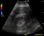
1) Endometrial mass: Endometrial mass is perhaps the most specific sign of RPOC. The figures below show a typical case of retained placenta. There is an echogenic mass of almost 8 x 5 cm inside the uterine cavity. Some fluid is seen along the margins of this mass.
2) Thickening of the endometrium: This is less specific and is less useful than seeing an endometrial mass.
3) Echogenic/complex fluid within the endometrial cavity. This is nonspecific and less sensitive than viewing an endometrial mass or thick endometrium and usually suggests hemorrhagic material.
Studies show that blood and hemorrhagic material within the uterine cavity can often mimic the appearances of RPOC.
Figure 1 Sagittal section of postpartum uterus. Large echogenic mass within the uterine cavity consistent with retained placenta.


Figure 2 Sagittal section of uterus; the retained placenta appears attached posteriorly to the myometrium.


Color Doppler Imaging of the Uterus
Color Doppler imaging can show the presence of or absence of flow within the uterine cavity. Absent flow is more suggestive of hemorrhagic material or clots in the uterus. However, nonviable placental tissue within the uterus can also show absent flow within the RPOC.
The color Doppler image of the retained placenta in Figure 3 clearly shows the difficulty in ruling out the presence of RPOC. Figures 1 and 2 are of a patient who delivered a healthy baby who was unable to deliver the placenta. She later began passing blood and debris through the vagina.
Other Factors that Indicate RPOC
RPOC may also be present when the following is visible:
• A clearly defined thin endometrial lining with minimal fluid within the endometrial cavity (normal appearance)
• A thin endometrial lining with echogenic material within the endometrial cavity that is separate from uterine wall (suggestive of blood clots)
• Echogenic material or a mass within the uterine cavity that is continuous with the uterine wall (indicates retained placenta)
Figure 3 Color Doppler of retained placental tissue. Note the absence of vascularity, which suggests nonviable placenta within the uterus.

In Figure 3, note that the uterine wall merges with the mass within the cavity, especially posteriorly. There is also a thinning of the myometrium posteriorly with some infiltration of the posterior wall of the uterus. This could indicate placenta accrete, which prevented the expulsion of the placenta.
Related Content
Images reproduced from Ultrasound-images.com View the video of this case
References:
References Â
De Vries, JI, van der Linden RM, van der Linden HC. Predictive value of sonographic examination to visualize retained placenta directly after birth at 16 to 28 weeks. JUM. 2000; 19: 7-12
Durfee SM, Frates MC, Luong A, et al. The sonographic and color Doppler features of retained products of conception. JUM. 2005;24:1181-1186.
Elsayes KM, Trout AT, Friedkin AM, et al. Imaging of the placenta: a multimodality pictoral review. Radiographics. 2009; 29:1371-1392.


S1E4: Dr. Kristina Adams-Waldorf: Pandemics, pathogens and perseverance
July 16th 2020This episode of Pap Talk by Contemporary OB/GYN features an interview with Dr. Kristina Adams-Waldorf, Professor in the Department of Obstetrics and Gynecology and Adjunct Professor in Global Health at the University of Washington (UW) School of Medicine in Seattle.
Listen