


Endometrial Polyps
As our ability to look inside the uterus improves, many women are told they have a common abnormality of the uterine lining, called endometrial polyps. An understanding of these common growths that develop inside the uterine cavity will help patients decide which course of treatment best suits them.
As our ability to look inside the uterus improves, many women are told they have a common abnormality of the uterine lining, called endometrial polyps. An understanding of these common growths that develop inside the uterine cavity will help patients decide which course of treatment best suits them.
The uterus is mostly composed of muscle. However, the inside lining of the uterus is made of "fluffy" endometrial tissue that grows and shrinks during the menstrual cycle. If a woman does not become pregnant, this lining sheds, causing a menstrual period. After a period, the lining grows rapidly under the influence of hormones like estrogen. Polyps are areas that grow a little too much. As they grow, they usually fan out but remain attached to a small stalk, kind of similar to a bush or a tree. The stalk is like the trunk of a tree, while the larger part of the polyp is like the branches (see photo below). They are usually about the size of a pencil eraser, although they can be even smaller. Rarely, polyps can grow to the size of an orange!

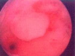

Photo taken during hysteroscopy of a small endometrial polyp. Notice the stalk.
Since most polyps are small, they probably do not often cause symptoms. However, when symptoms do occur, they usually include excessive bleeding during a menstrual period, or bleeding in between periods, or even spotting after intercourse. Some women report a few days of brown blood after a normal menstrual period. Polyps cause these symptoms because they dangle from their stalks and irritate the surrounding tissue, which causes the tissue to rub off, exposing tiny blood vessels. These blood vessels bleed, leading to spotting or vaginal bleeding. If the polyp interferes with the egg and sperm, it may make it hard to get pregnant. Nobody knows how common this is. It is also possible that they may lead to a slightly higher chance of miscarriage, but this is also unknown. Most gynecologists will remove polyps, as discussed below, if they are found in women with a history of miscarriage.
If a woman goes to her doctor complaining of spotting between periods or after intercourse, or very heavy bleeding during a menstrual period, her doctor will usually think of polyps as one of the many possible causes. Diagnosing endometrial polyps involves looking inside the uterine cavity. A regular ultrasound (also called a sonogram) usually does not diagnose polyps, because the pressure inside the uterus flattens the polyps, making them very hard to see. A special ultrasound, called a sonohysterogram (water ultrasound), allows doctors to see inside the uterus after a few drops of sterile water is carefully infused into the uterus through the vagina. The water opens the uterine cavity, allowing the doctor to see if any polyps are hanging around. Another diagnostic test is a hysterosalpingogram (HSG), which uses dye under pressure to open the uterus and tubes. A quick x-ray is then taken to see if any polyps are in the uterus. Finally, gynecologists are becoming more skilled at using the hysteroscope to look inside the uterus. This is a small, lighted tube that goes into the vagina then the uterus, to look around inside the uterus. Hysteroscopy using small tubes can be performed in the office, but larger tubes (used to remove large polyps or fibroids) usually require anesthesia in the hospital.


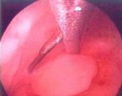
Tiny hysteroscopic scissors, about as big around as the ink tube on a standard writing pen, are used to cut the stalk.
If a polyp is diagnosed one of the first questions is "could this be cancer?" Fortunately, polyps only rarely turn cancerous. The risk does increase, but only slightly, as a patient passes age 50. The next question is often "how do you remove the polyp?" The old-fashioned way was to perform a D & C (dilatation and curettage). This involves a gentle scraping of the uterine lining. Unfortunately, this may miss the polyp completely, since this procedure is done solely by feel. Imagine a polyp dangling by a little stalk. As the scraping instrument goes by, it will likely just push the polyp out of the way without grabbing it. Thankfully, we now have hysteroscopes, which allow us to look right at the polyp as we grasp it or cut it away from the uterine lining. This ensures that the polyp (or, in some cases, multiple polyps) is removed. The photo below shows a polyp being removed by small scissors placed through a hysteroscope.
After removal of a polyp, the patient can return to work in a few days. She may notice a little spotting for a few days. Only a small percent of polyps seem to come back, but it is possible that months or years after treatment a polyp might recur. If you are diagnosed with endometrial polyps, please discuss treatment options with your doctor, who is in the best position to help you decide whether or not removal (called polypectomy) is necessary.
D. Ashley Hill, M.D.
Associate Director
Department of Obstetrics and Gynecology
Florida Hospital Family Practice Residency
Orlando, Florida


Reducing multifetal pregnancy through publicly funded IVF programs
April 26th 2024Learn how a mandatory elective single-embryo transfer policy in publicly funded in vitro fertilization programs significantly decreases multifetal pregnancy rates, offering insights into mitigating risks in assisted reproduction.
Read More

S1E4: Dr. Kristina Adams-Waldorf: Pandemics, pathogens and perseverance
July 16th 2020This episode of Pap Talk by Contemporary OB/GYN features an interview with Dr. Kristina Adams-Waldorf, Professor in the Department of Obstetrics and Gynecology and Adjunct Professor in Global Health at the University of Washington (UW) School of Medicine in Seattle.
Listen

Higher preterm birth risk found following cesarean delivery at full dilation
March 26th 2024Recent research highlights an association between cesarean delivery at full dilation and increased risk of subsequent preterm birth, prompting further investigation into childbirth practices and outcomes.
Read More