


Diagnosis and Laparotomic Treatment of an Interstitial Pregnancy
In a transvaginal coronal plane, you can see on the left-hand side of the fundus a flourishing pregnancy with an embryo of 9 weeks. In the last pictures of the clip a thin hypoechogenic endomiometrial layer is more easily seen.
I am a practicing OB-GYN, with 30 years of experience. In my experience, interstitial pregnancy is not as common as described in literature: 2-4% of all ectopic pregnancies. I submit a short video case of a suspected interstitial pregnancy at 9 weeks and welcome comments from the audience of this forum.
Best regards, Wolfgang
Please View the Video (1.6 mb)
In a transvaginal coronal plane, you can see on the left-hand side of the fundus a flourishing pregnancy with an embryo of 9 weeks. In the last pictures of the clip a thin hypoechogenic endomiometrial layer is more easily seen. The gestational sac is very eccentric. In none of the scans a connection of the chorion to the endometrial-decidual layer could be demonstrated. Although described in literature (Ackerman, Radiology 1993,189/1/83), I cannot recognize the "interstitial line sign".
The patient refused treatment with methotrexate and left the hospital saying that she was leaving for Serbia. She returned on Saturday night with pain in the left lower abdomen. On Sunday night the pain worsened and on Monday morning we decided to do a laparotomy for symptoms of haemoparitoneum. The patient had a previous laparotomy with salpingectomy for a left tubal pregnancy. On opening we found a moderate haemoperitoneum. The fundus uteri was covered with colon firmly attached to it. During accurate preparation and involuntary pressure on the fundus a 2.5 cm long fetus jumped out of the field. The thin layer of myometrium ruptured. All the chorion was accurately removed and the chorionic bed cleaned. The uterine opening was finally repaired with a continuous suture.
Please click on the images below for a larger view.


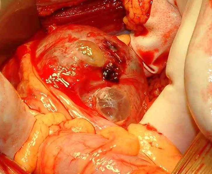
The enlarged left uterine angle after partial removal of the large intestine. The miometrium appears very thin and partially translucent. In the center the uterus is ruptured and the translucent corion is visible.

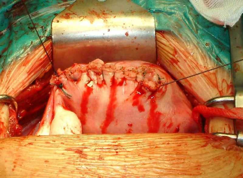

Repair of the uterus with a continous suture after removal of the pregnancy from the left uterine angle.


Reducing multifetal pregnancy through publicly funded IVF programs
April 26th 2024Learn how a mandatory elective single-embryo transfer policy in publicly funded in vitro fertilization programs significantly decreases multifetal pregnancy rates, offering insights into mitigating risks in assisted reproduction.
Read More

S1E4: Dr. Kristina Adams-Waldorf: Pandemics, pathogens and perseverance
July 16th 2020This episode of Pap Talk by Contemporary OB/GYN features an interview with Dr. Kristina Adams-Waldorf, Professor in the Department of Obstetrics and Gynecology and Adjunct Professor in Global Health at the University of Washington (UW) School of Medicine in Seattle.
Listen

Higher preterm birth risk found following cesarean delivery at full dilation
March 26th 2024Recent research highlights an association between cesarean delivery at full dilation and increased risk of subsequent preterm birth, prompting further investigation into childbirth practices and outcomes.
Read More