


3D imaging tracks fetal head and brain shape during labor
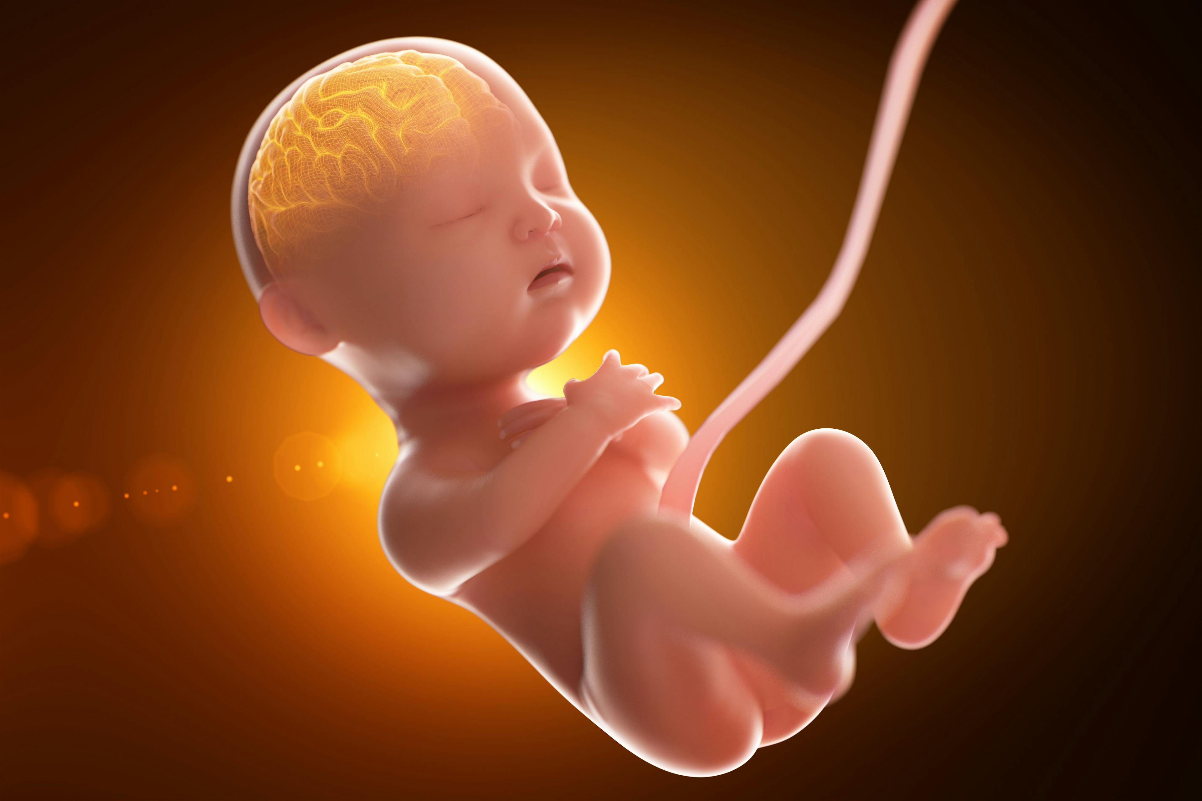
Recently published 3D MRI images show how the fetal head changes between prelabor and the second stage of labor to facilitate vaginal delivery.

©Connect world - stock.adobe.com

Even the most straightforward birth can be traumatic to an infant because its large head is not easily delivered, given the physiology of the maternal pelvis. Researchers from France have now published three-dimensional (3D) magnetic resonance imaging (MRI) images showing how the fetal head changes between prelabor and the second stage of labor to facilitate vaginal delivery.
Published in PLoSOne, the images are from seven women who were examined before going into labor using a 1 Tesla open field MRI and had another set of 3D MRI sequences done while they were in second-stage labor. Using the images, the authors created 3D reconstructions of each part of the fetal head to study fetal head molding and brain shape changes as labor progresses.
Patients were selected on the presumption that if their pregnancies were normal, the deliveries would be normal. However, two of the seven patients developed clinical cephalopelvic dystocia and required cesarean section for delivery.
All seven fetuses had fetal head molding with overlapping cranial sutures during the second stage of labor. Only two of the newborns, however, had deformed head contours, showing the flexibility of the molding system. The researchers said that overlapping of the cranial sutures was most evident in the anterior-posterior direction at the coronal and lambdoid suture. In all cases, at least one of the parietal bones was shifted to a position below the corresponding frontal bone. During the second stage of labor, fetuses had a significantly higher incidence of “sugarloaf” deformation of the brain and skull than before labor.
One of the major takeaways from their study, the authors said, lies in the fact that it raises the question of what defines “normal birth.” In this series, the child with the biggest skull deformation during the birthing process was born easily after only a few maternal efforts. That infant also showed signs of fetal distress despite continuous normal monitoring until the expulsion phase.
The researchers said that these occurrences indicate that the current definition (when a parturient gives birth by natural means with only a few maternal efforts) may not account for possible underlying issues, such as the ability of the fetal head to deform (compliance). The authors posited that the “normal birth” definition should be amended to “when a parturient gives birth naturally with only a few expulsive efforts and her child is fine and has no consequences due to the shape of its brain during the process.”


Reducing multifetal pregnancy through publicly funded IVF programs
April 26th 2024Learn how a mandatory elective single-embryo transfer policy in publicly funded in vitro fertilization programs significantly decreases multifetal pregnancy rates, offering insights into mitigating risks in assisted reproduction.
Read More

S1E4: Dr. Kristina Adams-Waldorf: Pandemics, pathogens and perseverance
July 16th 2020This episode of Pap Talk by Contemporary OB/GYN features an interview with Dr. Kristina Adams-Waldorf, Professor in the Department of Obstetrics and Gynecology and Adjunct Professor in Global Health at the University of Washington (UW) School of Medicine in Seattle.
Listen

Higher preterm birth risk found following cesarean delivery at full dilation
March 26th 2024Recent research highlights an association between cesarean delivery at full dilation and increased risk of subsequent preterm birth, prompting further investigation into childbirth practices and outcomes.
Read More